An egg contains approximately 2.0–2.2 grams of calcium — almost all of it in the shell. A laying hen producing 300 eggs over a 52-week cycle deposits more than 600 grams of calcium into eggshells. Her entire skeleton contains roughly 30 grams of calcium.
The arithmetic is immediate: no laying hen can sustain 300-egg production on skeletal calcium alone. She cannot produce it from feed calcium alone during the 16–18 hours it takes to form a shell overnight. She requires a supplementary calcium reserve — a specialized bone structure called medullary bone — that she builds during the rearing period and draws on during the dark phase of every laying cycle.
If that reserve is not built correctly before lay begins, the hen borrows calcium from structural bone instead. The result is osteoporosis, skeletal fractures, cage layer fatigue, reduced shell quality, and a laying trajectory that collapses before the flock reaches its economic potential.
Skeletal development in growing pullets is not a welfare consideration layered onto a production program. It is a foundational production input that determines the biological ceiling of everything that follows. And it is almost entirely a management outcome determined by what happens — or fails to happen — between week 1 and week 17.
The Skeleton a Laying Hen Needs: Two Types of Bone
Understanding how to build the right skeleton requires understanding that laying hens carry two functionally distinct types of bone simultaneously.
Cortical (Structural) Bone
Cortical bone is the dense, load-bearing bone that forms the shaft of long bones — the femur, tibiotarsus, and humerus — and provides the structural framework that allows the bird to stand, walk, perch, and access feeders and drinkers. It is built during the rearing period through a combination of adequate dietary calcium, phosphorus, vitamin D₃, and the mechanical stimulus of normal physical activity.
Once cortical bone is lost — whether through nutritional deficiency during rearing or calcium mobilization during intensive lay — it cannot be fully rebuilt while the bird is in production. The hen is laying simultaneously from the calcium stores she draws on and the dietary calcium she is consuming. There is no metabolic capacity to rebuild structural bone while producing 5–6 eggs per week.
This is why skeletal deficits accumulated during rearing are permanent production liabilities. They cannot be corrected by improving the laying ration after transfer.
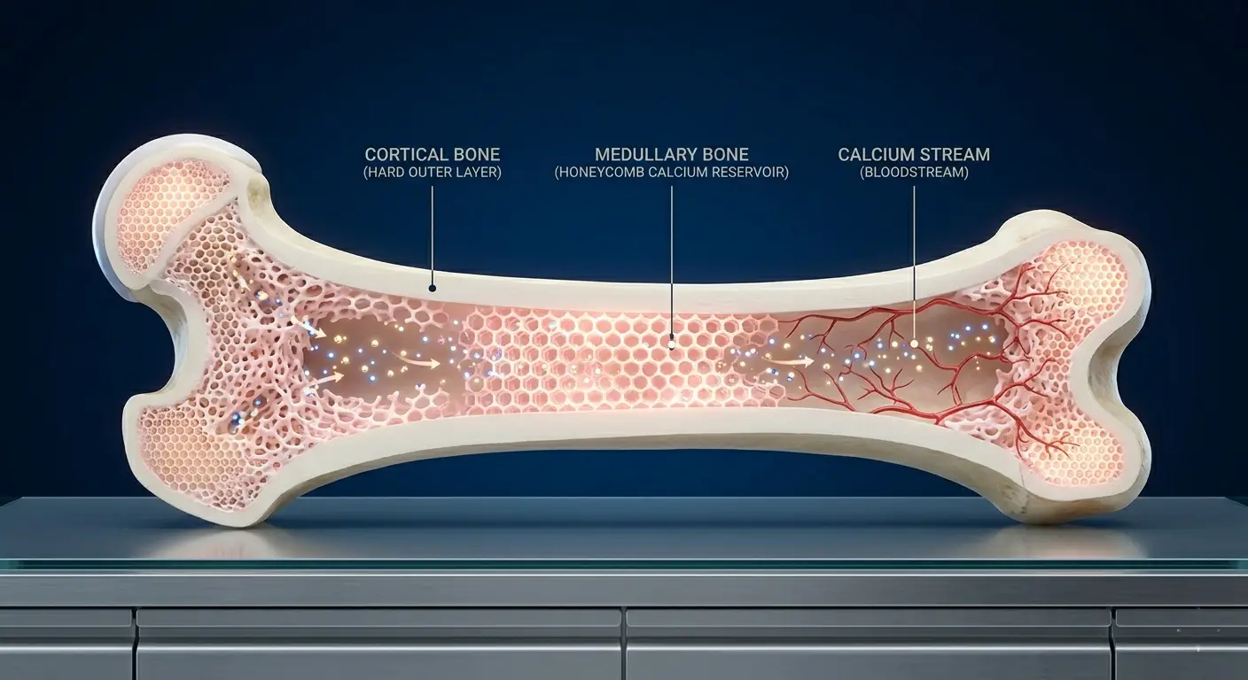
Medullary Bone
Medullary bone is a specialized, rapidly mobilizable calcium reserve that fills the medullary cavity of long bones — the hollow interior space. It develops exclusively in female birds under the influence of estrogen as they approach sexual maturity, typically beginning 10–14 days before the first egg is laid.
Medullary bone is biologically distinct from cortical bone: it is spongy in texture, highly vascularized, and can be deposited and resorbed within hours. During eggshell calcification — which occurs almost entirely during the dark period — the hen resorbs medullary bone calcium into the bloodstream and deposits it into the shell. During the following day, dietary calcium from feed rebuilds the medullary reserve in preparation for the next shell.
A hen with well-developed medullary bone entering lay has a calcium buffer that stabilizes shell quality even when dietary calcium absorption is temporarily incomplete. A hen with poorly developed medullary bone enters lay without that buffer and begins drawing on cortical bone within the first weeks of production.
The Calcium Timeline: What Must Happen and When
Skeletal development across the rearing period is not a linear process. Calcium demands and the skeleton’s capacity to respond to them change dramatically across the four phases of pullet development.
Phase 1: Foundation Building (Week 1–6)
During the starter phase, the priority is rapid skeletal growth — laying down cortical bone mass and establishing the frame size (measured by shank length) that the bird will carry into production. Dietary calcium at this stage is 0.90–1.00% of the ration, which is sufficient for growth without overloading the renal system.
The critical error during this phase is feeding a ration with excessive calcium, typically caused by using a laying ration or a calcium-boosted ration during the starter period. Calcium above 1.2% in the first six weeks causes calcium nephrosis: calcium deposits in the kidney tubules that permanently reduce renal function and calcium-processing capacity. A pullet with calcium-damaged kidneys carries that impairment into the laying cycle, limiting her ability to regulate blood calcium during shell formation.
Phosphorus during this phase is equally important. The calcium-to-phosphorus ratio in the starter ration should be 2:1 to 2.2:1 (available phosphorus basis). Available phosphorus targets: 0.45–0.50% of ration. Phosphorus deficiency during skeletal growth produces rickets — soft, deformed bones that cannot support body weight and do not provide an adequate structural base for medullary bone development later.
Vitamin D₃ is the metabolic linchpin. Without adequate vitamin D₃, dietary calcium and phosphorus cannot be absorbed from the intestine or deposited into bone, regardless of how much is provided in the ration. Target 2,000–3,000 IU/kg of vitamin D₃ in the starter ration for pullets reared indoors without natural sunlight exposure — the standard condition in enclosed commercial pullet houses.
Phase 2: Frame Consolidation (Week 7–12)
During the growth phase, linear skeletal growth slows as the frame approaches its genetic ceiling. The biological priority shifts from establishing frame size to consolidating bone density — mineralizing the cortical bone matrix that was laid down during the starter phase.
Dietary calcium remains at 0.90–1.00%. Phosphorus targets: 0.40–0.45% available phosphorus. The calcium-to-phosphorus ratio is critical during this phase — an imbalanced ratio (too much calcium relative to phosphorus, or vice versa) impairs bone mineralization even when absolute levels of both minerals are adequate.
This is the phase where feed restriction is most commonly misapplied in pullet production. Controlled feeding during the grower phase is appropriate and necessary to prevent excess fat deposition, but feed restriction that reduces total mineral intake below daily requirements prevents adequate bone mineralization. Calculate mineral intake per bird per day, not just the percentage of the ration. If total feed allocation is reduced, verify that mineral intake remains above the daily minimum requirement.
Daily calcium intake target during the grower phase: 800–900 mg per bird per day.
Body weight and shank length should be measured simultaneously at weeks 8 and 12 to assess skeletal frame development independent of body condition. A pullet at target body weight with short shank length is carrying the weight in fat. A pullet at target body weight with appropriate shank length has built the frame that the laying ration will sustain.
Shank length targets for commercial brown-egg layer breeds:
| Age (Weeks) | Target Shank Length (mm) |
|---|---|
| 6 | 72–78 |
| 8 | 82–88 |
| 10 | 90–96 |
| 12 | 96–102 |
| 16 | 98–108 |
Phase 3: Developer Phase and Calcium Restriction (Week 13–16)
The developer phase is the most misunderstood nutritional period in layer pullet production. It is characterized by intentionally low dietary calcium — 0.90–1.00%, the same as the starter and grower — and this is correct.
The reasoning: medullary bone development does not begin until estrogen levels rise as the bird approaches sexual maturity, typically week 17–18. Providing high calcium before medullary bone development begins has no productive benefit — the excess calcium that cannot be stored in medullary bone is excreted, placing a load on the renal system, or it is deposited in soft tissues, which impairs rather than supports later production.
Pullets fed a pre-lay ration (high calcium, 2.0%+) starting at week 14 or 15 — a common management error driven by the belief that “more calcium earlier is better” — show higher rates of urolithiasis (calcium deposits in the ureters), reduced feed intake from excess calcium palatability suppression, and, paradoxically, weaker shells in early lay than birds maintained on correct developer rations through week 16.
Phosphorus targets in the developer phase: 0.38–0.42% available phosphorus. Protein reduction to 14–15% prevents premature stimulation of the reproductive axis before the skeleton is structurally ready.
Phase 4: Pre-Lay Calcium Loading (Week 17–18)
The pre-lay phase is the most critical nutritional transition in the rearing program. It is the period during which medullary bone development is triggered by rising estrogen levels, and dietary calcium must be increased in coordination with this biological process.
The transition from developer ration (0.9–1.0% calcium) to pre-lay ration (2.0–2.5% calcium) should begin at week 17, or earlier if the flock shows clear signs of sexual maturity onset (reddening combs, wattle development, birds investigating nest box locations). Do not wait until the first egg is observed to increase calcium. By the time the first egg appears, medullary bone development has been underway for 10–14 days. If dietary calcium was not increased before that process began, the bird built her initial medullary bone reserve from cortical bone, beginning the laying cycle already in calcium deficit.
Pre-lay ration targets:
- Calcium: 2.0–2.5%
- Available phosphorus: 0.40–0.42%
- Crude protein: 17–18% (increased from developer to support reproductive tract development)
- Vitamin D₃: 3,000 IU/kg (increase from grower/developer level to maximize calcium absorption efficiency)
The pre-lay ration bridges the developer and laying rations without the metabolic shock of moving directly from 0.9% calcium to 3.8–4.2% calcium on the day of transfer. That direct jump, without the pre-lay transition, is a primary cause of production dips in the first 2–4 weeks of lay and is frequently misdiagnosed as a housing or lighting problem rather than a nutritional transition failure.
Calcium Sources: Not All Calcium Is Equal in Poultry Nutrition
The source of dietary calcium matters as much as the quantity, particularly during the laying period when the hen must absorb calcium rapidly and continuously.
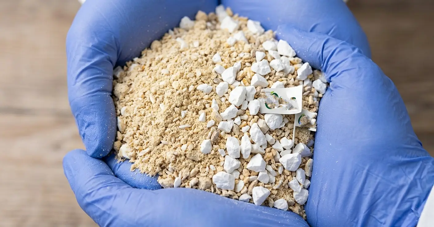
Limestone (Calcium Carbonate)
Limestone is the primary calcium source in virtually all commercial layer rations. Its effectiveness depends on particle size — a critical variable that is frequently overlooked in ration formulation.
Fine limestone (particle size < 0.5 mm) dissolves rapidly in the proventriculus and absorbs quickly. It is the appropriate form during the starter, grower, and developer phases when moderate, steady calcium absorption is the target.
Coarse limestone (particle size 2–4 mm, “oystershell grit” or “limestone chips”) dissolves slowly, releasing calcium over 8–12 hours. During the laying period, coarse limestone particles retained in the gizzard continue releasing calcium through the night — when the hen is calcifying the shell — reducing dependence on medullary bone resorption. Rations for laying hens should include 30–50% of total limestone in coarse particle form for this reason.
During the rearing period, fine limestone is adequate. Introducing coarse limestone at the pre-lay transition (week 17) — mixed at 50:50 with fine limestone — prepares the gut for the particle size mixture it will process throughout the laying cycle and improves the efficiency of shell calcification from the first egg onward.
Dicalcium Phosphate (DCP) and Monocalcium Phosphate (MCP)
These are the primary phosphorus sources in most commercial rations and also contribute calcium. The ratio of calcium-to-phosphorus in these sources must be accounted for in ration formulation — DCP provides approximately 21% calcium and 18% phosphorus. Overreliance on DCP as the calcium source in rations designed for high-calcium phases can create phosphorus excess — a different but equally damaging mineral imbalance.
Phytase Enzyme Supplementation
Most dietary phosphorus in plant-based feedstuffs — maize, soybean meal, wheat bran — is bound in phytate complexes that the hen’s digestive system cannot break down. Only 30–35% of total phosphorus in a maize-soybean meal ration is bioavailable without enzyme supplementation.
Adding microbial phytase to the ration at 500–750 FTU/kg increases phosphorus bioavailability by 0.10–0.15 percentage points — the equivalent of adding 1–2 kg of dicalcium phosphate per tonne of feed. At current DCP prices in Cameroon and Nigeria, phytase supplementation represents a significant feed cost reduction without compromising skeletal development. It also reduces phosphorus excretion in litter — a practical benefit in high-stocking-density operations where litter phosphorus accumulation accelerates ammonia production.
Vitamin D₃ and Its Downstream Roles
Vitamin D₃ is synthesized in the skin under ultraviolet light exposure. Pullets reared in enclosed commercial houses with no natural sunlight access are entirely dependent on dietary vitamin D₃ supplementation. In the high-UV environment of equatorial West Africa, open-sided houses with roof overhangs allow some ultraviolet penetration — but not reliably enough to be factored into ration formulation.
Vitamin D₃ is converted in the liver to 25-hydroxyvitamin D₃ (calcidiol), then in the kidney to 1,25-dihydroxyvitamin D₃ (calcitriol) — the biologically active form. Calcitriol increases intestinal calcium absorption by upregulating calcium transport proteins in the intestinal mucosa and stimulates renal calcium reabsorption, reducing urinary calcium loss.
Consequences of vitamin D₃ deficiency during rearing:
- Impaired calcium absorption regardless of dietary calcium level — the minerals are present, but cannot cross the intestinal wall at adequate rates
- Rickets in severe deficiency — soft, rubbery bones; reluctance to stand; characteristic “sitting on hocks” posture
- Reduced bone mineralization without frank rickets — a subclinical deficiency that only manifests at lay when calcium demand exceeds the hen’s limited absorption capacity
- Impaired immune function — vitamin D₃ receptors are present on immune cells and play a direct role in macrophage activation and pathogen response
Vitamin D₃ supplementation targets by phase:
| Phase | Dietary Vitamin D₃ Target |
|---|---|
| Starter (Week 1–6) | 2,000–3,000 IU/kg |
| Grower (Week 7–12) | 2,500–3,000 IU/kg |
| Developer (Week 13–16) | 2,500–3,000 IU/kg |
| Pre-lay (Week 17–18) | 3,000–3,500 IU/kg |
| Laying | 3,000–4,000 IU/kg |
Vitamin D₃ degrades during feed storage, particularly in hot and humid tropical environments where feed may sit in storage for 2–4 weeks before use. Add a 10–15% safety margin above the minimum target when formulating rations stored in non-climate-controlled conditions.
Diagnosing Skeletal Problems Before They Reach the Laying House
Skeletal deficiencies accumulated during rearing are visible before transfer if the right assessments are conducted. By the time a skeletal problem manifests as broken keel bones, cage layer fatigue, or shell quality decline in the laying house, the rearing window has closed, and the damage is irreversible.
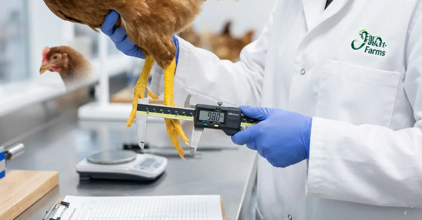
Shank Length Assessment
Shank length, measured from the hock joint to the base of the middle toe, is a breed-standardized skeletal frame indicator that is not affected by body condition or fat deposition. A bird with a short shank length for her age has a small skeletal frame — she will not develop adequate cortical or medullary bone mass to sustain high-rate production regardless of how well the laying ration is formulated.
Measure shank length in the same weighing sample used for uniformity assessment. Flag birds more than 5 mm below the breed standard at week 12 and 16 for nutrition review.
Keel Bone Palpation
The keel bone — the sternum — is the largest flat bone in the bird and the primary site of medullary bone deposition in laying hens. Palpating the keel in pre-transfer pullets reveals both skeletal development and body condition simultaneously.
A well-developed keel should be straight, firm, and have visible muscle coverage on both sides. A keel that is laterally deviated (curved to one side) indicates prior trauma or developmental abnormality. A keel with minimal muscle coverage (prominent to the touch with thin lateral coverage) indicates inadequate body condition despite adequate frame size.
Keel deviations detected at week 16 in more than 5% of sampled birds indicate a structural problem in the rearing environment — perch design, stocking density, or early nutritional deficiency — that must be investigated before the next flock cycle begins.
Tibia Breaking Strength (Flock-Level Audit)
In operations with access to basic laboratory equipment or veterinary extension services, tibia breaking strength measured from culled or dead birds provides a direct measure of cortical bone density. Target values for commercial pullet breeds at week 16: 180–220 Newtons.
Tibia breaking strength below 150 Newtons at week 16 indicates systemic bone mineralization failure that will manifest as high fracture rates at depopulation and reduced laying persistency throughout the cycle.
The Calcium-Phosphorus Imbalance: The Mistake That Undermines Everything Else
Every mineral interaction in skeletal development eventually traces back to the calcium-to-phosphorus ratio. A correct ratio with adequate absolute levels of both minerals produces sound bone. A correct ratio with deficient absolute levels produces inadequate bone volume. A correct absolute level with an incorrect ratio produces bone with structural or density abnormalities regardless of quantity.
The ratios that matter:
| Phase | Total Calcium | Available Phosphorus | Target Ca:P Ratio |
|---|---|---|---|
| Starter | 0.90–1.00% | 0.45–0.50% | 2.0–2.2:1 |
| Grower | 0.90–1.00% | 0.40–0.45% | 2.1–2.3:1 |
| Developer | 0.90–1.00% | 0.38–0.42% | 2.2–2.4:1 |
| Pre-lay | 2.00–2.50% | 0.40–0.42% | 5.0–6.0:1 |
| Laying | 3.80–4.20% | 0.38–0.42% | 9.0–11.0:1 |
The dramatic increase in calcium-to-phosphorus ratio between the developer and laying phases reflects the shift from skeletal building (where phosphorus demand is proportionally high) to shell calcification (where calcium demand dominates and phosphorus must be carefully limited to prevent urinary tract damage from excess).
Phosphorus excess during lay — caused by miscalibrated phytase, incorrect DCP inclusion, or insufficient phosphorus reduction when switching to laying ration — causes soft tissue calcium-phosphorus deposits and kidney damage that suppress calcium absorption and shell quality simultaneously.
Common Management Errors and Their Skeletal Consequences
Feeding laying ration to pullets before week 17. The 3.8–4.2% calcium in a laying ration is nephrotoxic to pullets whose renal calcium-processing capacity is not yet developed. It causes calcium nephrosis, permanently reduces renal function, and impairs calcium regulation throughout the laying cycle.
Delaying the pre-lay ration transition until the first egg. Medullary bone development begins 10–14 days before the first egg. A hen that receives 0.9% calcium while building her medullary reserve draws on cortical bone to do it. She enters life already in a structural calcium deficit.
Restricting feed during the grower phase without adjusting mineral density. Cutting feed allocation by 10% without increasing mineral concentration by 10% produces a 10% mineral deficit every day the restriction continues.
Ignoring phosphorus when calcium is increased. Increasing dietary calcium to “improve shells” without simultaneously reviewing phosphorus levels disrupts the ratio and impairs both bone mineralization and renal function.
Sourcing limestone without verifying particle size. Fine and coarse limestone look identical in a feed bag. Without particle size verification from the supplier, the slow-release calcium benefit of coarse limestone in the pre-lay and laying rations may not exist in practice.
Development and Calcium Loading in Growers
The skeleton of a laying hen carried into the laying house was built between week 1 and week 17. The medullary bone reserve she draws on for every egg she lays for the next 52 weeks was built in the 10–14 days before her first egg. Neither can be rebuilt once the laying cycle begins.
Getting skeletal development right requires the correct calcium level at each phase — low during the starter and grower periods to protect renal function, low during the developer period to avoid premature deposition before medullary capacity exists, and precisely timed during the pre-lay transition to build medullary reserve before the first shell demands it. It requires an accurate calcium-to-phosphorus ratio at every phase, adequate vitamin D₃ to make dietary calcium biologically accessible, and regular skeletal assessment through shank length measurement and body condition scoring throughout the rearing period.
The hen that lays 320 eggs in 52 weeks did not get that number from the laying house. She earned it in the rearing phase — one correctly mineralized bone at a time.