Disease is the third highest cost in commercial catfish production after feed and labor — and unlike feed and labor, disease costs are highly variable and potentially catastrophic. A well-managed catfish operation with good water quality, appropriate stocking density, and effective biosecurity may spend XAF 800,000–1,500,000 per year on routine health management for a 50-tonne operation. The same operation experiencing an uncontrolled Aeromonas outbreak, a Columnaris epizootic during a heat wave, or a viral hemorrhagic event without rapid diagnosis and response may lose XAF 5,000,000–20,000,000 in a single production cycle from mortality, treatment costs, and production delays.
The difference between controlled disease costs and catastrophic disease losses is almost entirely a management difference — specifically, the combination of preventive practices that reduce the probability of disease introduction and the diagnostic capability that identifies disease events early enough for effective intervention. A catfish farmer who recognizes Aeromonas hydrophila ulcers when they first appear on two or three fish in a tank and initiates treatment within 24 hours will typically lose 2–5% of that tank. A farmer who notices the same initial signs, attributes them to “normal” minor injuries, and returns three days later to find 30% of the tank dead has allowed the same infection to progress to the point where treatment is expensive, slow, and partially ineffective.
This guide covers the bacterial and viral diseases that most significantly affect commercial African catfish production in West and Central Africa — their clinical presentation, diagnostic approach, treatment protocols, and the water quality and management conditions that predispose fish to each infection.
The Relationship Between Water Quality and Bacterial Disease
Why Most Bacterial Catfish Diseases Are Secondary to Stress
The single most important concept in catfish disease management is that the majority of bacterial infections affecting commercial production are caused by opportunistic pathogens — bacteria that are present in virtually every aquatic environment at low levels, including the most carefully managed catfish tanks, but that only cause disease when the fish’s immune defenses are compromised by stress.
Aeromonas hydrophila is present in essentially every body of water where catfish are farmed. Flavobacterium columnare (the Columnaris pathogen) is similarly ubiquitous. The fish’s intact immune system — mucus barrier, skin integrity, innate immune cells, and adaptive immune responses — holds these bacteria at bay under normal conditions. When the fish’s immune defenses are compromised by:
- Dissolved oxygen below 4 mg/L (even transiently)
- Ammonia or nitrite above stress thresholds
- Temperature outside the optimal range (particularly above 32°C or below 18°C)
- Physical injury from handling, crowding, or abrasion
- Nutritional deficiency (vitamin C, vitamin E, essential fatty acids)
- Prior viral infection that depletes lymphocytes
…the bacteria that were being successfully excluded suddenly find an undefended entry point and establish an infection that escalates rapidly once initiated.
The management implication: Treating an active bacterial infection while the predisposing stress remains in place typically produces partial and temporary improvement followed by recurrence. Effective bacterial disease management requires simultaneously treating the infection and correcting the predisposing stress condition — otherwise the fish that survive the antibiotic treatment course immediately re-establish the same stress state and become re-infected.
Aeromonas hydrophila — Motile Aeromonad Septicemia (MAS)
Clinical Presentation
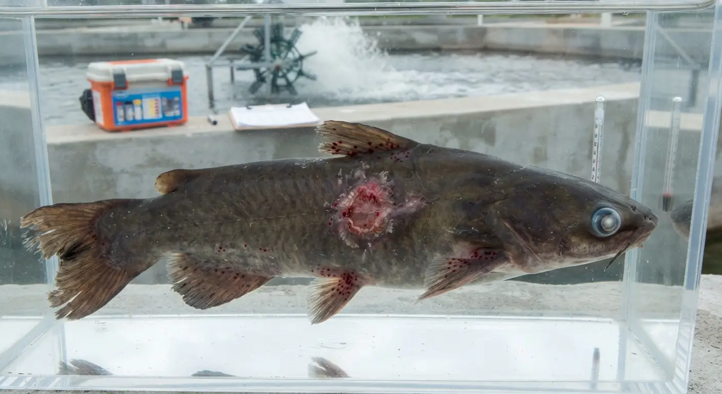
Aeromonas hydrophila and related motile Aeromonas species (A. sobria, A. veronii) are the most common causes of bacterial disease in African catfish across West and Central Africa. The clinical syndrome they produce — Motile Aeromonad Septicemia (MAS) — is a systemic bacterial infection that can range from mild skin ulceration to acute septicemic death within 24–48 hours of clinical signs.
External signs (early to moderate infection):
- Ulcers and skin lesions: Circular or oval areas of skin erosion, typically 0.5–3.0 cm in diameter, exposing underlying red or hemorrhagic muscle tissue. Ulcers can appear anywhere on the body surface but are particularly common at the base of fins, on the flank, and at the head. Fresh ulcers have a bright red, moist appearance; older ulcers may develop gray or white necrotic margins.
- Hemorrhages at fin bases: Redness and petechial (pinpoint) hemorrhage at the base of dorsal, pectoral, and caudal fins — one of the earliest external signs, sometimes appearing before frank ulceration develops.
- Fin erosion: Progressive destruction of fin tissue beginning at the fin margins, giving fins a ragged, frayed appearance. Particularly common in crowded tanks where fin damage from physical contact initiates infection sites.
- Exophthalmia (pop-eye): Unilateral or bilateral protrusion of one or both eyes from the orbital socket, produced by the accumulation of inflammatory fluid behind the eye. The protruding eye is often hazy or opaque.
- Abdominal distension: In severe systemic infection, accumulation of bloody or clear fluid in the abdominal cavity produces visible distension. Fish with abdominal distension typically show the disease in an advanced stage and have a poorer prognosis.
Behavioral signs:
- Reduced feeding response — often the first sign visible in a tank before external lesions are obvious
- Lethargy — affected fish rest on the tank bottom or near the surface rather than swimming actively
- Loss of equilibrium in advanced cases — fish unable to maintain normal position, listing or spiraling
Acute septicemic form: In water quality crashes or post-transport stress events, MAS can present as acute mortality without prominent skin lesions — fish simply die rapidly without the progressive ulceration that characterizes the more typical subacute presentation. Post-mortem examination typically reveals petechial hemorrhage on internal organs and congested, hemorrhagic liver tissue.
Diagnosis
Clinical diagnosis: The combination of ulcers, fin erosion, and hemorrhage at fin bases in the context of a water quality event, high stocking density, or recent handling stress is highly suggestive of MAS and sufficient for initiating empirical treatment while formal laboratory confirmation is sought.
Laboratory confirmation (where available):
- Bacterial culture from the margin of a fresh lesion (not the center of an old ulcer, which typically contains secondary contaminants rather than the primary pathogen) using blood agar or tryptone soya agar at 28°C
- Biochemical characterization or API identification of recovered colonies
- Antibiotic sensitivity testing (antibiogram) — essential for guiding effective antibiotic selection given the increasing prevalence of antibiotic-resistant Aeromonas strains in operations that have used antibiotics repeatedly without culture-and-sensitivity guidance
Treatment Protocol
Step 1: Address the predisposing stress immediately
Before or simultaneously with antibiotic treatment, correct the water quality or management condition that predisposed the fish to infection:
- If DO is below 5 mg/L: increase aeration immediately
- If TAN or nitrite is elevated: increase water exchange or add salt (for nitrite) as described in the water quality article
- If temperature is above 32°C: add shade or cooler water exchange
- If stocking density is excessive: consider an emergency partial harvest to reduce biomass
Step 2: Salt treatment (supportive, immediate)
Common salt (NaCl) at 2–5 g/L in the tank water serves multiple roles in MAS management:
- Reduces osmotic stress on fish with damaged skin (ulcers create areas where osmotic balance is disrupted)
- Reduces nitrite toxicity (if nitrite is elevated — the competitive inhibition mechanism described in the water quality article)
- Has mild bacteriostatic properties against some Gram-negative bacteria at higher concentrations
Apply as a bath or continuous tank treatment — for continuous treatment, 2–3 g/L is appropriate; for a short-term bath (1–2 hours), 5–10 g/L is used outside the tank.
Step 3: Antibiotic treatment
Oxytetracycline (OTC): The most widely used antibiotic for MAS in African catfish production in West Africa — broad-spectrum tetracycline with documented efficacy against Aeromonas spp. Available as water-soluble powder for bath/tank treatment or incorporated into medicated feed.
- Water bath treatment: 10–20 mg/L oxytetracycline in tank water for 1 hour, then exchange with clean water; repeat every 48 hours for 3–5 days
- Medicated feed: 55–83 mg OTC per kg body weight per day in feed, for 10 consecutive days
Florfenicol: A broad-spectrum antibiotic with excellent activity against Aeromonas and generally good tissue penetration — available as a premix for incorporation into medicated feed at 10–15 mg/kg body weight per day for 10 days. More expensive than OTC but increasingly preferred where OTC resistance has been documented.
Amoxicillin: Effective against sensitive strains; available in some markets as a water-soluble powder for bath or medicated feed incorporation.
Critical antibiotic management principles:
Complete the course: Do not discontinue antibiotic treatment when clinical signs improve — the full course (10 days for feed-incorporated antibiotics) must be completed to prevent the selection of partially resistant survivors that become the dominant strain in subsequent outbreaks.
Observe withdrawal periods: All antibiotics have a mandatory withdrawal period before fish are sold for human consumption — oxytetracycline typically 21–28 days, florfenicol typically 12–28 days depending on temperature. Track treatment dates for each tank and do not harvest treated fish before the withdrawal period has elapsed.
Rotate classes with each new outbreak: Do not use the same antibiotic class for consecutive outbreaks in the same operation without sensitivity testing — repeated use of the same antibiotic in the same population selects for resistance. Rotate between OTC, florfenicol, and other available classes.
Culture and sensitivity first where possible: Where a diagnostic laboratory is accessible within 24–48 hours, submit samples for bacterial culture and antibiogram before initiating antibiotic treatment, or at the same time as initiating empirical treatment. The sensitivity result guides adjustment of the treatment if the empirical choice is not fully effective.
Prevention
- Maintain DO above 5 mg/L continuously — the single most effective MAS prevention measure
- Handle fish with minimal physical trauma — use smooth, knotless nets; avoid crowding during harvest or transfer
- Minimize stocking density at the peak heat season when immune competence is lowest
- Administer vitamin C supplementation at 500–1,000 mg/kg in feed to support wound healing and immune function
- Vaccinate broodstock against Aeromonas where commercial vaccines are available (intraperitoneal injection vaccination of broodstock confers partial maternal antibody protection in larvae)
Flavobacterium columnare — Columnaris Disease
What Makes Columnaris Distinctive
Columnaris disease, caused by the Gram-negative rod Flavobacterium columnare, is the second most significant bacterial pathogen of commercial African catfish after Aeromonas — and arguably the most acutely dangerous, because it can cause mass mortality within 24–48 hours under favorable conditions without the gradual ulceration progression that provides warning time in Aeromonas infections.
Flavobacterium columnare is a uniquely temperature-sensitive pathogen — its virulence increases dramatically with water temperature above 28°C, and epidemic outbreaks are strongly associated with heat events. An operation that experiences a water temperature spike to 32–34°C during a hot dry season period and simultaneously has any of the predisposing stress factors (low DO, physical injury, overcrowding) faces acute Columnaris risk.
Clinical Signs
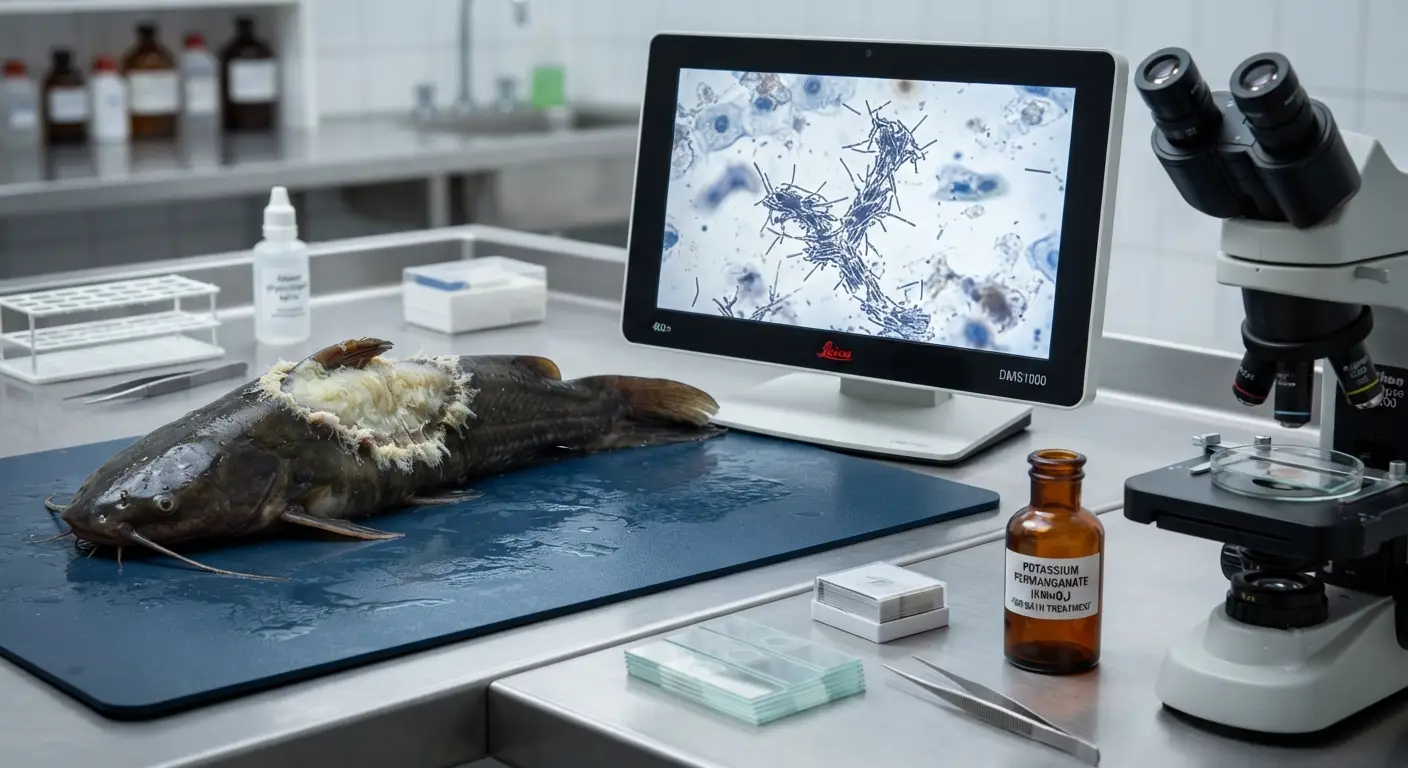
The saddle-back lesion: The characteristic early lesion of Columnaris — a pale gray to yellowish-white area of skin erosion and necrosis on the dorsal surface of the body, extending from near the dorsal fin outward and downward on both sides, creating a “saddle” appearance. The lesion has a distinctive frayed, cotton-wool texture from the bacterial growth and necrotic tissue at its margin.
Gill infection (gill rot): Columnaris very frequently infects gill tissue — in some outbreaks, gill infection precedes visible skin lesions. Infected gills show areas of necrosis (pale, brown, or whitish patches on the gill filaments), increased mucus production, and in severe cases, fusion or erosion of the gill lamellae. Fish with severe gill Columnaris show severe respiratory distress — rapid opercular movement, surface breathing, and behavioral DO stress even at adequate water DO — because the gill tissue damage prevents normal oxygen extraction.
Fin erosion: Similar in appearance to MAS fin erosion, but the Columnaris lesion often has a more yellowish discoloration and a distinctive musty odor from bacterial metabolites.
Oral and facial infection: Lesions on the lip, jaw, and around the mouth — “mouth rot” — are a characteristic Columnaris presentation, particularly in fingerlings.
Diagnosis
Columnaris can be distinguished from MAS by:
- The characteristic saddle-back lesion morphology (not typical of MAS)
- The cotton-wool texture of lesion margins (the bacterial growth of Flavobacterium produces this texture; Aeromonas ulcers have cleaner margins)
- Wet mount microscopy from the lesion margin: long, slender, gliding rods that aggregate in rotating “haystacks” — a distinctive microscopically visible behavior of Flavobacterium that can be observed with a standard light microscope
Treatment Protocol
Potassium permanganate (KMnO₄): The most effective and most practical treatment for external Columnaris in commercial catfish operations — potassium permanganate is a strong oxidizing agent that rapidly kills Flavobacterium on external surfaces at concentrations that fish tolerate:
- Indefinite bath treatment: 2 mg/L in tank water, maintained continuously. This is the concentration at which Flavobacterium is killed while most catfish tolerate the treatment for extended periods. Monitor fish closely during treatment — at concentrations above 4 mg/L, oxygen is consumed by the oxidizing reaction and fish may show respiratory stress.
- Short-term bath: 10 mg/L for 30–60 minutes outside the production tank, then return fish to clean water.
Oxytetracycline (medicated feed): 55–83 mg/kg body weight per day for 10 days — addresses systemic infection when Flavobacterium has invaded beyond the skin surface into deeper tissues.
Copper sulfate: 0.5–1.0 mg/L in tank water for Columnaris treatment — effective but pH-dependent (effectiveness decreases above pH 8.0) and potentially toxic to fish above 1.0 mg/L. Use with caution and precise dosing.
Critical additional response — temperature reduction:
Because Columnaris virulence is strongly temperature-dependent, reducing water temperature during an outbreak — by increasing the proportion of cooler supply water, providing shade, or reducing stocking density to lower metabolic heat — is a direct management intervention that reduces bacterial growth rate and virulence simultaneously with drug treatment. An outbreak at 32°C that is treated with potassium permanganate while temperature remains at 32°C will respond more slowly than the same outbreak treated while temperature is brought to 28°C.
Edwardsiella tarda — Edwardsiellosis
Clinical Presentation
Edwardsiella tarda causes a systemic infection characterized by:
- Gas-filled cysts or abscesses in the muscle tissue — the characteristic “gas disease” of Edwardsiellosis, producing visible bubble-like cavities in the musculature visible at post-mortem
- External ulcers similar to MAS but with a distinctive foul odor from the gas-producing metabolic activity of E. tarda
- Systemic signs: lethargy, loss of equilibrium, hemorrhagic liver at post-mortem
Edwardsiellosis is less common than MAS or Columnaris in West African commercial catfish but occurs sporadically and can cause significant mortality in stressed populations, particularly where organic loading of ponds or tanks is high.
Treatment
Oxytetracycline (55–83 mg/kg/day in medicated feed for 10 days) is effective against sensitive strains. Trimethoprim-sulfonamide combinations have shown efficacy where OTC resistance is present.
Streptococcal Infections
Streptococcus and Lactococcus Infections in Catfish
Streptococcal bacteria — particularly Streptococcus agalactiae and Lactococcus garvieae — cause a specific neurological disease syndrome in African catfish characterized by:
- Erratic, spiral, or “corkscrew” swimming behavior — the pathognomonic clinical sign of bacterial meningitis in fish
- Loss of equilibrium — fish unable to maintain normal dorsal-up orientation
- Exophthalmia (pop-eye) — bilateral protrusion of both eyes in many cases
- Hemorrhage at the base of fins and around the eyes
Streptococcal infections in catfish in West Africa are associated with high water temperature (above 30°C), high stocking density, and periods of immunological stress. They are particularly significant in tilapia production across the region but are documented in African catfish as well.
Treatment
Florfenicol (10–15 mg/kg body weight per day in medicated feed for 10 days) and amoxicillin are the antibiotics with documented efficacy against Streptococcus in fish. OTC has limited efficacy against Gram-positive bacteria including Streptococcus — empirical OTC treatment of a suspected Streptococcal infection is often ineffective, reinforcing the importance of culture and sensitivity testing.
Viral Infections
The Fundamental Difference Between Viral and Bacterial Disease Management
Viral infections require a fundamentally different management approach than bacterial infections — there are no antiviral medications available for commercial fish production. The management options for viral disease are:
- Prevention through biosecurity: Excluding the virus from entering the production population through strict quarantine and biosecurity protocols
- Supportive care: Optimizing all other conditions (water quality, nutrition, reduced stress) to support the fish’s own immune response to the viral infection
- Vaccination: Where commercial vaccines are available for the specific virus (limited in current African catfish commercial production)
- Culling: In severe outbreaks of highly pathogenic viruses, depopulation and disinfection may be the only way to stop spread and allow restocking
Catfish Fry Virus (Channel Catfish Virus — CCV, Ictalurid Herpesvirus 1)
Relevance to African catfish production: CCV (Ictalurid Herpesvirus 1) primarily affects North American Ictalurus punctatus (channel catfish) — it is not currently documented as a significant pathogen of Clarias gariepinus in West African production. However, it is mentioned here because operations that import channel catfish genetics or fingerlings from North American sources face CCV introduction risk.
Catfish Rhabdovirus
Several rhabdovirus species have been isolated from African catfish (Clarias gariepinus) in Europe and Africa, causing hemorrhagic disease with:
- Severe hemorrhage on internal organs and skin
- Rapid mortality in juvenile fish
- Explosive outbreak pattern (high morbidity within days)
Management: Strictly biosecurity-based — no treatment is available. Outbreaks require depopulation, thorough disinfection with appropriate virucides (sodium hypochlorite 200–500 ppm contact time 30 minutes for wet surfaces), and extended fallow period before restocking from a known virus-free source.
Iridoviral Infections
Several iridoviruses have been documented in African catfish in Asian production systems (where Clarias is extensively farmed), causing systemic infection with:
- Spleen enlargement (splenomegaly) — often the most prominent gross pathological finding
- Hemorrhage at multiple sites
- High mortality in juvenile fish
No specific treatment; management is biosecurity and population-level response to confirmed outbreaks.
The Viral-Bacterial Co-infection Problem
In commercial catfish production, viral and bacterial infections frequently co-occur — a viral infection that suppresses immune function creates the opportunity for the opportunistic bacteria (Aeromonas, Columnaris) that are always present in the environment to establish secondary infections. This co-infection pattern means that:
- The visible clinical signs of the disease are often primarily those of the bacterial secondary infection (ulcers, hemorrhage, fin erosion) even though a primary viral infection initiated the immune collapse
- Antibiotic treatment addresses the visible bacterial component but not the underlying viral primary infection
- Mortality continues despite antibiotic treatment because the viral pathogen continues to cause immune suppression
Diagnostic indicator of possible viral-bacterial co-infection: Antibiotic treatment that produces partial improvement followed by recurrence, or that controls visible lesions but does not stop mortality, suggests a primary viral component that is not addressed by the antibiotic. Where laboratory PCR testing for relevant viruses is accessible, testing concurrent with or after the antibiotic trial provides definitive differentiation.
The Post-Mortality Investigation Protocol
Why Every Significant Mortality Event Requires Investigation
As established in the pig series health management guidance — and applying equally to fish health — every mortality event that exceeds the background mortality rate (above 0.5% per day in grow-out, above 1% per week in fingerlings) should be investigated rather than attributed to vague “stress” or “water quality” without specific diagnosis.
The investigation sequence for a catfish disease event:
Step 1: Water quality assessment (same day)
- Measure DO, temperature, TAN, NO₂, pH
- Document any recent water quality event (pump failure, power failure, feed spike, algal bloom crash)
- Correct any water quality parameter outside target range immediately
Step 2: Clinical observation (same day)
- Examine 10–20 sick and recently dead fish for external signs
- Categorize the lesion pattern: ulcers/hemorrhage (MAS), saddle-back/cotton-wool (Columnaris), spiral swimming (Streptococcal/neurological), sudden death without lesions (acute septicemia or viral)
- Note: Are lesions bilateral or unilateral? Localized or generalized? Present on specific body regions?
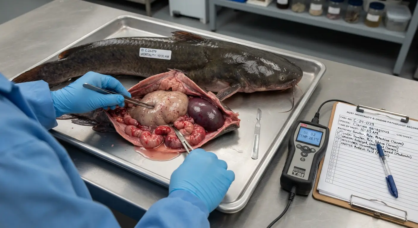
Step 3: Post-mortem examination (same day, on freshly dead fish)
- Examine: liver color and texture (pale, friable, or hemorrhagic?), spleen (enlarged?), kidney (swollen, pale, or hemorrhagic?), abdominal fluid (present? clear or bloody?), gas pockets in muscle?
- Document findings
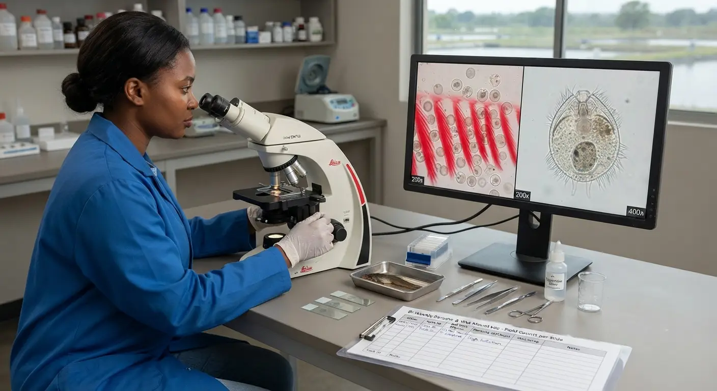
Step 4: Wet mount microscopy (if microscope available)
- Gill wet mount: parasites (Trichodina, Ichthyophthirius, Gyrodactylus), gill necrosis
- Skin/lesion margin wet mount: Flavobacterium columnare characteristic gliding rods
- Blood smear: abnormal blood cells, bacteria in bloodstream
Step 5: Laboratory submission (within 24–48 hours)
- Submit 3–5 freshly dead or moribund (not already dead) fish on ice for bacterial culture, sensitivity testing, and histopathology
- Provide history: tank parameters, feeding rate, recent events, clinical signs timeline
Step 6: Treatment based on diagnosis (not before)
- Initiate appropriate treatment based on the specific diagnosis or most supported empirical diagnosis
- Document: treatment product, dose, duration, tank identity, number of fish treated, withdrawal period end date
Summary
Bacterial infections are the dominant disease challenge in commercial African catfish production — and their management is fundamentally a water quality management problem with a treatment component, not a treatment problem with a water quality component. Aeromonas hydrophila and Flavobacterium columnare account for the majority of significant disease mortality across West and Central African commercial catfish operations, and both are opportunistic pathogens whose virulence depends on the fish’s immune competence being compromised by one or more of the identifiable stress factors that correct water quality management prevents.
Early diagnosis — recognizing the characteristic clinical presentations of each major pathogen and initiating investigation before the outbreak has consumed significant fish biomass — is the difference between a manageable production loss and a catastrophic one. The investigation protocol in this guide converts a mortality observation from an observable fact into actionable diagnostic information within hours rather than days, allowing treatment to be specific and effective rather than empirical and delayed.
Viral infections add a management dimension that antibiotics cannot address — the prevention through biosecurity focus, the supportive care approach, and the recognition that bacterial co-infections secondary to viral primary events require both antibiotic treatment and ongoing investigation of the underlying cause that antibiotics will not resolve.
The next article addresses parasitic and fungal disease management — the second tier of catfish health challenges that frequently co-occurs with the bacterial infections covered here.