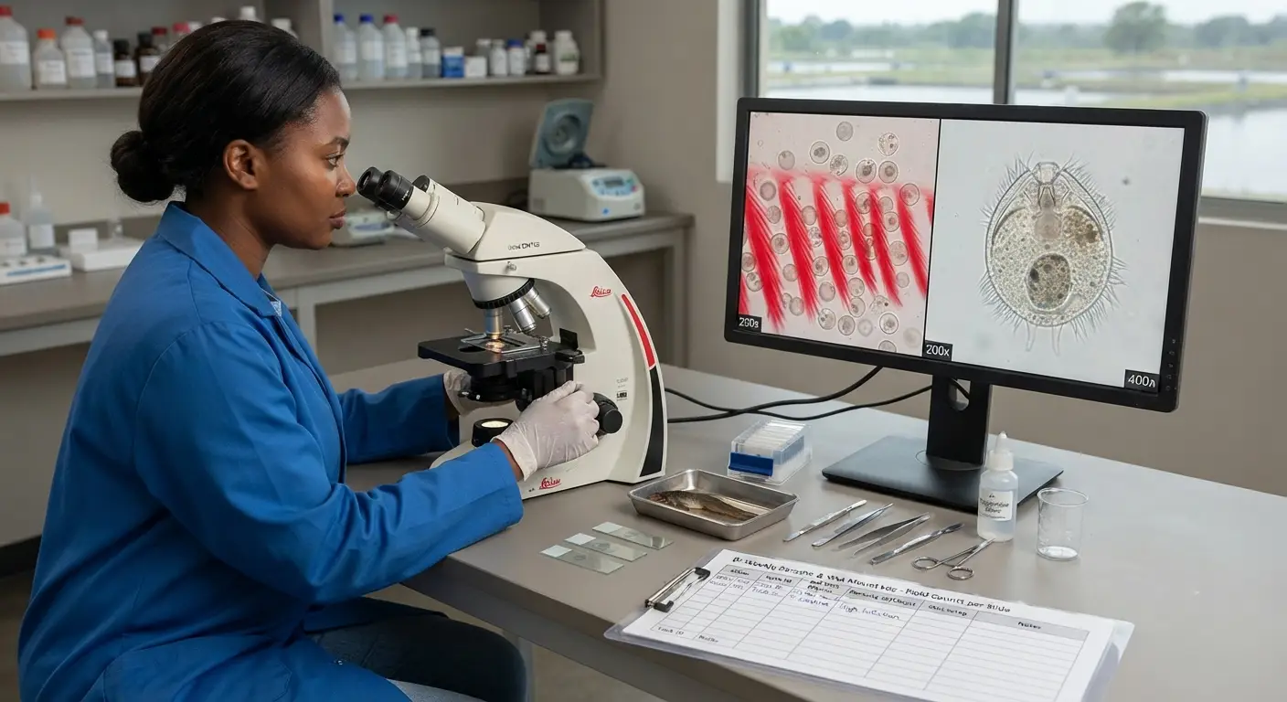
The catfish farmer who walks through the grow-out facility and sees fish that are feeding, swimming, and appearing externally normal concludes that the operation is healthy. A parasitologist examining gill wet mounts from those same fish under a microscope would frequently find Trichodina at moderate to high infestation levels, Gyrodactylus on the skin surface, and perhaps early Ichthyophthirius cysts establishing in the gill epithelium — none of which is visible to casual external observation, all of which is actively consuming resources that would otherwise support fish growth and immune function.
Parasites are the most consistently underdiagnosed production challenge in commercial catfish farming precisely because their effects are chronic and distributed rather than acute and concentrated. A tank with moderate Trichodina infestation does not experience an obvious disease event — it simply underperforms. FCR is 15–20% worse than in an equivalent uninfested tank. Daily growth rate is 10–15% lower. Immune function is sufficiently compromised that the next bacterial challenge — an Aeromonas outbreak following a water quality event — causes significantly higher mortality than it would in a parasite-free population. The connection between the chronic parasitic infestation and the elevated bacterial disease susceptibility is rarely identified without the active diagnostic monitoring that most commercial operations do not systematically implement.
This guide covers every significant parasitic and fungal disease of commercial African catfish production — the specific pathogens, their clinical presentations, the diagnostic methods for identifying them, and the treatment and prevention protocols that remove them from production systems before their cumulative impact on growth, FCR, and disease susceptibility translates into measurable production losses.
Ectoparasites — External Parasites on Skin and Gills
Trichodina — The Most Prevalent External Parasite
What Trichodina is: A ciliated protozoan parasite — a single-celled organism approximately 30–100 μm in diameter with a characteristic disc-like structure bearing a ring of denticles (hook-like structures) on its ventral surface that it uses to anchor to host tissue. Trichodina species are obligate parasites of fish skin and gills — they cannot survive independently in the water for extended periods and must transfer between fish hosts to complete their life cycle.
Life cycle: Trichodina reproduces by binary fission directly on the host fish — no intermediate host is required. Population growth on a single fish can be rapid under favorable conditions: high stocking density (facilitating fish-to-fish contact transmission), warm temperatures (accelerating protozoan replication), and poor water quality (compromising the host’s immune defenses). Infestation levels on a single fish can reach thousands of individuals per square centimeter of gill tissue in severe cases.
Clinical signs:
Mild infestation: No visible external signs detectable by casual observation — fish appear healthy and continue feeding. The only detectable evidence of mild Trichodina infestation is reduced growth rate and slightly elevated FCR compared to expected benchmarks, and susceptibility to secondary bacterial infection following any immune challenge.
Moderate infestation: Increased mucus production visible as a slightly milky or cloudy appearance to the skin surface; minor behavioral changes including some flashing behavior (brief rolling or rubbing movements as if trying to scratch the skin against objects); possibly mildly elevated surface breathing frequency.
Severe infestation: Heavy mucus production creating a visible bluish-white discoloration of the skin; pronounced flashing behavior; significant respiratory distress as gill tissue is damaged by heavy infestation; reduced feed intake; increasing lethargy; secondary bacterial infection of skin areas where mucus barrier has been disrupted.
Diagnosis:
The definitive diagnosis of Trichodina requires microscopy — no external clinical sign is specific enough to distinguish Trichodina from other causes of skin and gill irritation.
Wet mount preparation:
- Anesthetize or euthanize 3–5 representative fish (ideally moribund individuals showing signs, or randomly selected from the population being assessed)
- Using a clean coverslip or blunt scalpel, scrape mucus from the skin surface (flank, dorsal area) and place the scraping in a drop of tank water on a glass slide
- Remove one gill arch, clip a section of gill filament, and prepare a wet mount in tank water on a separate slide
- Examine under 100× to 400× magnification — Trichodina appear as disc-shaped organisms with the characteristic denticle ring, moving in a distinctive rotating and gliding motion
Population assessment: Count the number of Trichodina per microscopic field at 100× magnification from 5–10 fields on each slide:
- Below 5 per field: low infestation — monitor but treatment not immediately required
- 5–20 per field: moderate infestation — treatment indicated
- Above 20 per field: heavy infestation — urgent treatment required
Treatment:
Salt treatment (sodium chloride): The most accessible and most practically effective treatment for Trichodina in West and Central African commercial catfish operations:
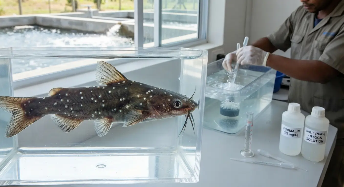
- Long-term (24–48 hours): 3–5 g/L NaCl continuously in tank or pond water — osmotic stress at this concentration disrupts protozoan membrane function while fish tolerate it well for this duration
- Short bath (30–60 minutes): 10–15 g/L NaCl outside the production system, followed by return to clean water — more stressful for fish but effective for severe infestations or where continuous tank treatment is not feasible
Formalin bath: 150–250 mg/L formalin (37% formaldehyde solution) for 30–60 minutes — effective against Trichodina but requires oxygen monitoring during treatment (formalin consumes dissolved oxygen) and good ventilation (formaldehyde vapors are hazardous to the treatment operator)
- Alternatively: 25–50 mg/L formalin continuously in tank or pond water for 24 hours
Potassium permanganate: 2–5 mg/L in tank water for 60 minutes — effective against external protozoa including Trichodina; less potentially hazardous than formalin
Post-treatment verification: Repeat microscopy 48–72 hours after treatment completion to confirm parasite clearance — a treatment that reduces infestation from severe to moderate has not achieved control and requires either a second treatment course or investigation of whether the source of re-infestation (new fish, water source) is ongoing.
Ichthyophthirius multifiliis — White Spot Disease (“Ich”)
What makes Ich different from other external parasites: Ichthyophthirius multifiliis (Ich) has a life cycle with a free-living stage that makes it uniquely difficult to treat with in-tank chemical treatments — the parasite spends a portion of its life cycle encysted in fish tissue (the trophont stage, where it is protected from external chemical treatment) and a portion as a free-swimming infective stage (the theront) that is chemical-sensitive. Treatment must be timed to the chemical-sensitive free-living stage.
Life cycle at 28°C:
- Trophont: The feeding stage — the parasite burrows under the skin or gill epithelium, grows, feeds on host tissue, and becomes the white spot (1–2 mm diameter) visible to the naked eye. Duration: 4–7 days at 28°C. TREATMENT DOES NOT REACH THE TROPHONT STAGE — it is protected by host tissue.
- Tomont: After the fully developed trophont exits the fish, it settles on the tank bottom and encysts, dividing internally to produce hundreds of infective theronts. Duration: 12–18 hours at 28°C.
- Theront: The infective free-swimming stage that seeks a host fish and penetrates the skin or gill epithelium to begin a new trophont stage. Duration: 4–8 hours before death if a host is not found. TREATMENT IS EFFECTIVE AGAINST THERONTS.
Clinical signs:
- White spots (1–2 mm) on the skin surface and fins — visible to the naked eye without microscopy in moderate to heavy infestation; the characteristic “white spot” appearance is pathognomonic
- Intense flashing behavior as fish attempt to relieve the irritation of trophonts burrowing in the skin
- Heavy surface breathing and clustering near water inlets as gill infestation reduces oxygen extraction capacity
- Rapid deterioration — in a susceptible naive population at high stocking density, Ich can cause significant mortality within 5–7 days of first visible signs
Treatment strategy — treating the tank environment, not the fish directly:
Since trophonts in fish tissue are inaccessible to chemical treatment, effective Ich control requires eliminating theronts in the water environment — preventing re-infestation of fish as trophonts mature and exit, and breaking the cycle until all trophonts have exited and the fish population is no longer a host reservoir.
Formalin treatment (most effective): 25 mg/L continuously in tank water for 5–7 days, or 150–250 mg/L bath for 60 minutes every 48 hours for 3 treatments — the continuous low-dose approach is preferred as it maintains lethal theront exposure throughout the cycle duration
Salt treatment: 3–5 g/L NaCl continuously for 5–7 days — less effective than formalin against Ich but safer for fish; suitable for mild infestations or as a complement to formalin treatment
Temperature elevation: Raising water temperature to 30–32°C (within African catfish’s tolerance range) accelerates the Ich life cycle, shortening the trophont stage and reducing the duration of the infective period — complements chemical treatment by reducing the total treatment duration needed
Prevention:
Ich is most commonly introduced through new fish (fingerlings or broodstock) from infested sources. Standard quarantine with prophylactic formalin or salt treatment of all incoming fish before release to production tanks is the most effective prevention. Ich theronts do not survive more than 4–8 hours without a host — a tank that has been depopulated and left fallow for 24 hours at 28°C (48 hours at lower temperatures) is effectively Ich-free.
Gyrodactylus — Monogenean Skin Flukes
What they are: Gyrodactylus species are small (0.5–1.0 mm) monogenean flatworms that live on the skin surface and fins of fish, attaching with a posterior holdfast organ equipped with hooks and anchors. They are viviparous — they give birth to live juveniles directly on the host fish, allowing rapid population growth without a free-living stage.
Clinical signs:
Gyrodactylus causes skin and fin irritation similar to Trichodina — increased mucus production, flashing behavior, fin erosion at margins. Heavy infestation produces similar external signs to Trichodina. Microscopy of skin scrapings at 100× magnification shows the characteristic elongated flatworm body with the posterior attachment organ bearing central hooks visible at 200–400×.
Treatment:
Gyrodactylus is eliminated by the same salt, formalin, and potassium permanganate treatments effective against Trichodina — though the monogenean’s more robust body requires the higher concentrations in the effective range:
- Formalin bath: 200–250 mg/L for 60 minutes
- Salt: 5–10 g/L for 30 minutes (short bath outside production tank)
- Praziquantel: 2–10 mg/L bath for 1–3 hours — highly effective against monogeneans with minimal fish toxicity; more expensive than formalin or salt but preferred where available
Dactylogyrus — Gill Flukes
What they are: Related to Gyrodactylus but oviparous (egg-laying) and primarily inhabiting the gill epithelium rather than the skin surface. Dactylogyrus infestation (gill flukes) produces gill inflammation, excess mucus on gill surfaces, and compromised respiratory efficiency similar to Columnaris gill disease — differentiation requires microscopy of gill tissue wet mounts.
Clinical signs: Respiratory distress disproportionate to the measured dissolved oxygen level — fish breathing rapidly or surfacing despite adequate DO in the water. Gill wet mount microscopy shows the flatworm body with anterior hamuli (hooks) visible at 100–200×.
Treatment: Same protocol as Gyrodactylus — formalin bath, praziquantel, or potassium permanganate.
Internal Parasites
Intestinal Worms (Nematodes and Cestodes)
Internal helminth parasites — roundworms (nematodes) and tapeworms (cestodes) — infest the intestinal tract of African catfish, particularly fish sourced from wild populations or from ponds with access to invertebrate intermediate hosts. Their effects in commercial production are:
- Competition with the fish for absorbed nutrients — reducing the effective nutrient availability from the consumed diet and directly worsening FCR
- Physical irritation of the intestinal mucosa — reducing absorptive efficiency and increasing susceptibility to enteric bacterial infection
- In heavy infestations: intestinal blockage or rupture (rare but documented in severe cases)
Diagnosis: Post-mortem examination of the intestinal tract — uncoiling and examining the intestine under running water reveals adult worms. Fecal egg flotation examination (the same McMaster technique described in the pig series parasite article) quantifies egg output as a population-level indicator.
Treatment:
Levamisole: 50–100 mg/kg body weight administered in medicated feed for 3 days — effective against nematode intestinal parasites
Praziquantel (for cestodes): 50–100 mg/kg body weight in medicated feed for 5 days — specific activity against tapeworms and monogenean flukes; limited activity against nematodes
Prevention: Fish sourced from wild populations or earthen ponds with natural invertebrate communities should be dewormed during quarantine before introduction to production systems. All fingerlings entering the grow-out system from external sources benefit from a prophylactic deworming treatment as part of the stocking protocol.
Acanthocephala (Spiny-Headed Worms)
Acanthocephala species infest the intestine, attaching with a spiny retractile proboscis that penetrates the intestinal wall — causing more severe tissue damage than smooth-bodied nematodes. The intermediate host for most Acanthocephala species is a crustacean (amphipod, isopod, or similar) — infestation risk is higher in fish sourced from wild environments or earthen ponds where these invertebrates are present.
Clinical signs: Intestinal perforation (in severe cases), peritonitis, and progressive emaciation. At post-mortem: worms visible in the intestine with the characteristic spiny proboscis embedded in the intestinal wall.
Treatment: Praziquantel in medicated feed at 50–100 mg/kg body weight for 5 days has partial efficacy against Acanthocephala; complete elimination of heavy infestations may require repeated treatment courses.
Fungal Infections
Saprolegnia — Water Mold
What Saprolegnia is: Not a true fungus taxonomically, but historically classified and managed as one — Saprolegnia species are oomycetes (water molds) that are ubiquitous in freshwater environments. They are primarily saprophytic (decomposing dead organic matter) but will infect living fish tissue that has been damaged by prior injury, disease, or stress — making Saprolegnia almost always a secondary infection rather than a primary pathogen.
The sequence of events in Saprolegnia infection:
- Predisposing event creates damaged tissue — a bacterial ulcer from MAS, a physical injury from handling, a Ich lesion, a spawn injury during the stripping procedure
- Saprolegnia zoospores, present in the water, land on the damaged tissue and establish hyphal growth
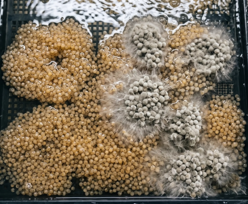
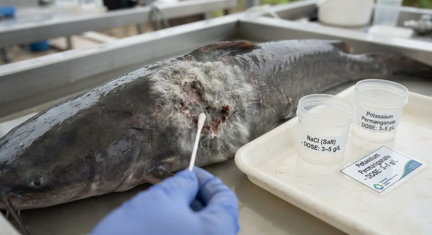
- The cotton-wool-like white to grayish fungal growth (the visible mass of hyphae) extends outward from the initial infection site across the damaged tissue surface
- The expanding mycelium consumes damaged tissue and can extend into healthy adjacent tissue, particularly at water temperatures below 20°C where the host immune response to fungal infection is weakest
Clinical signs:
The cotton-wool-like fuzzy white to grayish-brown growth on the skin, fins, or around eggs in the hatchery is pathognomonic for Saprolegnia — no other catfish pathogen produces this appearance. Location is typically at:
- Previously ulcerated or damaged skin areas
- Fin bases that have been eroded by bacterial infection or physical damage
- Wounds from spawning handling procedures on broodstock
- Around the head region in stressed fish
- On catfish eggs in the incubator (Saprolegnia on eggs can spread to adjacent healthy eggs, significantly reducing hatch rate)
Treatment:
Salt treatment: 2–5 g/L NaCl in tank water for 24–48 hours — osmotic stress at this concentration inhibits Saprolegnia hyphal growth and reduces active lesions. The most practical treatment for production tanks given salt’s broad-spectrum benefit against multiple concurrent problems.
Potassium permanganate: 2–5 mg/L for 60–120 minutes bath treatment — oxidizes the fungal hyphae effectively. Repeat every 48 hours for 3 treatments.
Hydrogen peroxide: 1,000–1,500 mg/L for 30 minutes bath treatment outside the production system — highly effective against Saprolegnia with low fish toxicity at this concentration and exposure time; becoming more widely available and used as the historically used malachite green (now banned in food fish production in most jurisdictions) is replaced
Malachite green: Historically the most effective Saprolegnia treatment — now banned for use in food fish production in most jurisdictions due to carcinogenic concerns. Do not use malachite green in fish destined for human consumption.
Prevention:
Saprolegnia is a secondary pathogen — preventing Saprolegnia infection requires preventing the primary conditions that create damaged tissue:
- Effective management of bacterial ulcer disease (MAS) reduces the ulcerated tissue that provides Saprolegnia entry points
- Careful handling to minimize physical trauma
- Maintaining water quality above the stress thresholds that compromise the host immune defense against Saprolegnia establishment
- In the hatchery: formalin treatment of eggs (50–100 mg/L for 15 minutes daily during incubation) prevents Saprolegnia establishment on egg batches with partial fertility
Environmental Stress Diseases
Nutritional Muscular Dystrophy (Vitamin E / Selenium Deficiency)
As established in the nutritional requirements article, vitamin E and selenium deficiency produce oxidative damage to muscle tissue, presenting as:
- Pale, white-streaked skeletal muscle visible in fresh fish or at post-mortem
- Reduced swimming vigor and apparent muscle weakness
- Elevated mortality in the affected population
Differentiation from bacterial myositis (bacterial muscle infection): Nutritional muscular dystrophy shows diffuse, bilaterally symmetric pale streaking visible on both sides of the fish; bacterial myositis typically shows localized lesions without the diffuse symmetry of nutritional deficiency.
Treatment/correction: Correct the feed formulation to include adequate vitamin E (60–200 mg/kg feed) and selenium (0.5–1.5 mg/kg feed) in stabilized forms. Injectable vitamin E/selenium combinations (as described in the pig series first-aid kit article — the same products used for pigs are sometimes used for fish in emergency intervention situations) can provide rapid correction in severely affected fish.
Gas Bubble Disease (Dissolved Gas Supersaturation)
What causes it: Water supersaturated with dissolved gases — most commonly nitrogen — produces gas emboli (bubbles) in fish blood vessels and tissues when the fish moves from high-pressure (deep water) to lower-pressure zones, or when supersaturated water delivers gas directly into the circulatory system. Causes include:
- Water supply from a pump that draws air into the suction line, producing nitrogen-supersaturated supply water
- Water supply from a borehole where groundwater is supersaturated with dissolved nitrogen from geological sources
- Cascade aeration systems that overaerate above 120% saturation
Clinical signs:
- Gas bubbles visible under the skin, particularly in the fins and around the head
- Exophthalmia (pop-eye) from gas accumulation behind the eye
- Rapid mortality in severe cases
Diagnosis and treatment: Measure dissolved gas saturation with a dissolved gas meter (a supersaturation meter, not a standard DO meter) — above 110% total dissolved gas is the threshold for gas bubble disease risk. Treatment is aeration management — degassing the incoming water supply before it enters the fish tanks. A cascade degasser or simple aeration structure that allows gas exchange with the atmosphere reduces supersaturation to safe levels.
Thermal Shock Mortality
Rapid temperature change — particularly rapid drops from cooling water addition, rainy season onset, or shade removal exposing a previously shaded tank — can cause acute mortality even when the final temperature is within the safe range.
Vulnerable situations:
- Replacing a significant fraction of tank water with supply water that is more than 5°C cooler
- Heavy rainfall events into open earthen ponds (cool rainwater rapidly mixing with warm pond water)
- Moving fish from an indoor warm holding tank to outdoor cooler tanks without temperature acclimatization
Prevention: The same acclimation principles described in the fingerling transport article — never change fish water temperature by more than 2–3°C per hour. Temper replacement water to within 1–2°C of the tank temperature before large water exchange events.
Monitoring for Parasites — The Routine Microscopy Protocol
Why Routine Microscopy Is Non-Negotiable
The single most effective health management upgrade available to most West African commercial catfish operations at modest cost is the implementation of routine wet mount microscopy — examination of skin and gill samples from production fish under a light microscope on a scheduled basis.
The cost of a basic laboratory microscope appropriate for this application: XAF 150,000–500,000 (USD 250–833). The cost of not doing routine microscopy: chronic Trichodina infestation reducing FCR by 15–20% across the entire production cycle, undetected early Ich infestation before it becomes a tank-wide outbreak, and elevated bacterial disease susceptibility from the immune compromise of undiagnosed external parasite loads.
Routine Microscopy Protocol
Sampling frequency:
- Every 2 weeks in fingerling tanks (highest risk period)
- Monthly in grow-out tanks (maintenance monitoring)
- Before and after any new fish introduction
- Whenever FCR rises above target without an identified water quality or nutritional cause
- At the first sign of flashing behavior, skin discoloration, or respiratory distress
Sampling procedure:
- Anesthetize 5–8 randomly selected fish from the tank (clove oil at 50–80 mg/L induces anesthesia in 1–3 minutes; remove fish to fresh water after examination)
- Prepare gill wet mount: clip a small section of gill filament, place in a drop of tank water on a glass slide, coverslip
- Prepare skin wet mount: scrape mucus from the flank with a clean coverslip edge, mix in a drop of tank water on a slide, coverslip
- Examine under 100× for Trichodina, Ichthyophthirius trophonts, and monogenean flukes; 200–400× for Trichodina denticle detail and parasite identification confirmation
Recording the result:
| Date | Tank | Fish sampled | Gill result | Skin result | Action taken |
|---|---|---|---|---|---|
| Date | Tank ID | Number | Parasite species + infestation level | Parasite species + infestation level | Treatment initiated or monitoring continued |
Integrating Microscopy Findings With Production Performance Data
The most diagnostically powerful use of routine microscopy data is correlating parasite infestation levels with concurrent FCR data from the same tanks. A tank showing elevated FCR without obvious water quality or feed quality explanation — and concurrently showing moderate Trichodina or Gyrodactylus on wet mount examination — has identified the cause of the FCR deviation. Treatment that reduces parasite infestation, followed by monitoring of FCR trend, confirms the causal relationship if FCR returns toward target following parasite control.
This correlation — parasite level × production performance impact — builds the farm-specific database that quantifies the financial cost of parasite infestation in that specific operation and makes the economic case for the routine monitoring investment that prevents infestation from reaching production-significant levels.
Treatment Safety — Protecting Fish, Farm Staff, and the Food Chain
Handling Chemical Treatments Safely
Several treatments used in catfish disease management — formalin, potassium permanganate, copper sulfate, praziquantel — require specific safety precautions for the person applying them:
Formalin: Formaldehyde vapors are carcinogenic and respiratory irritants. Always handle formalin outdoors or in well-ventilated areas. Use chemical-resistant gloves and avoid skin contact. Do not inhale vapors during preparation or application.
Potassium permanganate: Strong oxidizer that causes skin and eye irritation. Use chemical-resistant gloves. Store away from organic materials that could ignite on contact.
Salt: Safe to handle — no specific precautions beyond normal hygiene.
Praziquantel: Low acute toxicity; standard hygiene precautions appropriate.
Withdrawal Periods for Chemical Treatments
Food safety critical principle: Any chemical applied to food fish production systems may leave residues in fish tissue that pose a food safety risk if fish are harvested before the withdrawal period has elapsed.
Known withdrawal periods for commonly used catfish treatments:
- Formalin (formaldehyde): No established withdrawal period in most jurisdictions where it is registered for aquaculture; follow local regulatory guidance
- Salt: No withdrawal period — sodium chloride residue at treatment levels is not a food safety concern
- Potassium permanganate: No significant tissue residue concern at treatment concentrations; typical guideline is 48–72 hours pre-harvest clearance
- Praziquantel: Withdrawal period of 14–21 days in most markets where guidelines exist
- Oxytetracycline: 21–28 days (temperature and dose dependent)
- Florfenicol: 12–28 days
Record treatment events and calculate clearance dates for every tank receiving any chemical treatment, and verify the clearance date before harvest. The same record-keeping discipline described in the pig series treatment log applies directly here — the withdrawal period tracking function of the treatment record is a food safety requirement, not merely a production management tool.
Summary
Parasites, fungal infections, and environmental stress diseases form the second tier of catfish health challenges — less immediately dramatic than the acute bacterial infections covered in the previous article but equally significant to production economics when they operate chronically at subclinical infestation levels.
The management framework for these conditions parallels the approach established throughout this series: systematic monitoring (routine wet mount microscopy on a defined schedule) provides early detection before infestation reaches production-impacting levels; specific diagnosis guides specific treatment rather than empirical broad-spectrum application; correct treatment with verified-effective agents at appropriate doses and durations achieves infestation clearance rather than partial suppression; and predisposing stress correction prevents re-establishment of the same infestation following treatment.
The farm that implements routine monthly wet mount microscopy and responds to identified parasite infestations with appropriate treatment will consistently achieve better FCR than the farm whose parasite management is reactive — responding only to visible clinical signs that by definition indicate the infestation has already reached production-impacting levels. The microscope is the most financially productive equipment purchase available to most commercial catfish operations — not because it cures anything, but because it sees what casual observation cannot.
The next article covers farm biosecurity, quarantine protocols, and predator control — the prevention layer that keeps all of these pathogens from entering the production system in the first place.