The egg that cracks into a hot pan tells the buyer everything they need to know about the eggs they will buy next week. If the yolk stands tall and the albumen grips the pan without spreading into a thin watery film, the buyer notes the quality and reorders. If the yolk breaks immediately, the albumen spreads flat, and the whole egg looks old and oxidized before the heat touches it, the buyer remembers that too — and makes a different choice next week.
Internal egg quality is the consumer experience behind the brand promise. The shell communicates quality before purchase. The interior confirms it — or fails to — at the moment of use. For institutional buyers who are serving eggs to guests, interior quality failure is not merely a quality complaint. It is an operational problem that disrupts plated presentation, undermines food service standards, and ends supply relationships.
Haugh units — the standardized measurement of albumen gel strength and height — are the scientific language of that consumer experience. A Haugh unit score above 72 is Grade AA albumen. A score of 60–72 is Grade A. Below 60 is Grade B. The score tells exactly where on the freshness and quality spectrum a given egg sits. And unlike the external quality variables that can be managed at collection and storage, the Haugh unit at the moment of laying is determined by the hen’s nutritional status, age, disease history, and the specific proteins she deposited in the albumen during oviduct transit — inputs that must be managed weeks before the consumer cracks the egg.
This article covers the biology of albumen and yolk quality, the factors that determine Haugh unit scores and yolk strength at laying, the measurement protocols that provide actionable data, and the nutritional and management interventions that maintain Grade AA internal quality through the laying cycle.
The Biology of Internal Egg Quality
Albumen Structure and Haugh Units
The albumen (egg white) is not a uniform liquid. It consists of four distinct layers: an outer thin albumen, a layer of thick albumen (the chalaziferous layer), the chalazae — the twisted fibrous cords that anchor the yolk at the center — and an inner thin albumen layer immediately surrounding the yolk.
The thick albumen layer constitutes approximately 57% of total albumen volume and is responsible for the gel-like standing height that Haugh units measure. It is a glycoprotein matrix — primarily ovomucin — that forms a three-dimensional network maintaining the albumen in its characteristic standing structure.
Ovomucin is the protein most directly responsible for thick albumen gel integrity. It is a high-molecular-weight glycoprotein that forms fibrous strands, creating the structural lattice of thick albumen. As the egg ages, the pH rises (from CO₂ loss as discussed in the storage article), and at pH above 8.5, ovomucin undergoes a conformational change — the fibrous strands dissociate, the gel structure collapses, and the previously standing thick albumen becomes a spreading thin liquid.
This pH-mediated ovomucin dissociation is the primary mechanism of Haugh unit decline over time — and it is also the mechanism by which heat stress accelerates quality decline. At elevated ambient temperatures, CO₂ loss is faster, pH rises more rapidly, and ovomucin dissociation occurs earlier in the egg’s post-lay life.
What Haugh Units Actually Measure
The Haugh unit (HU) calculation was developed by Raymond Haugh in 1937 as a standardized measure of albumen quality. The measurement combines:
- The height of the thick albumen at a defined distance (10mm) from the edge of the yolk, measured in millimeters using a micrometer
- The weight of the egg is used to adjust for the mathematical relationship between egg size and albumen height
The formula: HU = 100 × log(H − 1.7 × W^0.37 + 7.6)
Where H = albumen height in mm and W = egg weight in grams.
In practice, Haugh unit measurement requires a calibrated micrometer stand or an automatic Haugh unit meter (available from egg quality laboratory suppliers). For farms without this equipment, a practical approximation is visual estimation of albumen spread:
- Grade AA equivalent (HU above 72): Albumen stands visibly above the yolk when broken onto a flat plate; the edge of the thick albumen is clearly defined with a distinct boundary from the spreading thin albumen; total spread is less than 8 cm from the center
- Grade A equivalent (HU 60–72): Albumen somewhat spread; thick and thin albumen less clearly differentiated; total spread 8–11 cm
- Grade B equivalent (HU below 60): Albumen thin and watery; no distinct thick layer; total spread exceeding 11 cm
For farms supplying institutional buyers who specify Grade AA, weekly visual assessment of 20–30 break-out samples provides the quality monitoring data that guides both storage management and nutritional interventions.
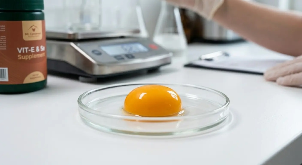
Yolk Quality: The Vitelline Membrane
The yolk is surrounded by a thin protein membrane — the vitelline membrane — that maintains its spherical shape and separation from the albumen. Vitelline membrane strength determines the yolk index: the ratio of yolk height to yolk width when the egg is broken out onto a flat surface. A fresh, strong-membraned yolk has a high yolk index (0.42–0.45); an aging or weak-membraned yolk flattens to a low index (below 0.30).
The biology of vitelline membrane weakness:
The vitelline membrane is composed primarily of glycoproteins (ovomucin-related) deposited in the oocyte during follicular development in the ovary. The membrane degrades through enzymatic action (proteolysis) and physical oxidative damage after laying, at rates governed by the same temperature and time relationship that affects albumen quality.
However, vitelline membrane strength at the moment of laying — before any post-lay degradation has occurred — varies with the hen’s nutritional status during follicular development. Specific nutrients known to affect vitelline membrane integrity:
Vitamin E (tocopherol): As the primary fat-soluble antioxidant in the oocyte, vitamin E protects the vitelline membrane’s lipid components from oxidative damage during the 7–10 days of rapid yolk growth preceding ovulation. Vitamin E deficiency or insufficiency at suboptimal levels produces fragile vitelline membranes that rupture more easily at break-out, even in fresh eggs.
Selenium (selenoprotein activity): Selenium-dependent glutathione peroxidase neutralizes lipid peroxides that would otherwise damage vitelline membrane protein structures. Insufficient selenium amplifies oxidative damage during follicular development.
Riboflavin (Vitamin B2): A cofactor for the oxidative defense enzymes in the oocyte. Riboflavin deficiency produces a suite of egg quality deficiencies, including weak vitelline membranes and increased blood spot incidence.
The Factors That Determine Internal Quality at Laying
Factor 1: Age of the Hen
Internal egg quality at laying declines progressively as the hen ages — an unavoidable biological trend. The mechanism is progressive deterioration of the oviduct epithelium that secretes albumen proteins, combined with declining secretory activity of the magnum tubular glands that produce ovomucin.
The practical trajectory:
| Laying Age | Expected Haugh Unit Range | Expected Yolk Index |
|---|---|---|
| Week 1–4 of lay (early) | 82–92 HU | 0.42–0.46 |
| Week 10–20 (developing) | 78–88 HU | 0.40–0.44 |
| Week 25–40 (peak and early mid) | 74–84 HU | 0.38–0.42 |
| Week 45–60 (mid to late) | 68–80 HU | 0.35–0.40 |
| Week 60–72 (late lay) | 62–75 HU | 0.32–0.37 |
This age-related decline is not linear — it accelerates in the final 15–20 weeks of the laying cycle. The practical implication: a farm supplying institutional buyers who specify Grade AA quality (HU above 72) can expect to maintain that specification reliably through week 50–55, then faces increasing difficulty in the final 15–20 weeks unless storage optimization and nutritional support are applied.
Factor 2: Heat Stress
Heat stress during the laying period affects albumen quality through two mechanisms:
Reduced oviduct transit time: At ambient temperatures above 30°C, the hen’s metabolic rate and physiological stress responses alter oviduct motility — the contractile movement that moves the forming egg from the infundibulum through the magnum (where albumen is added) to the isthmus, uterus, and vagina. Accelerated transit reduces the time available for magnum secretory cells to deposit full albumen protein volume, resulting in thinner albumen with lower Haugh unit at laying.
Reduced ovomucin secretion: The magnum tubular glands that secrete ovomucin are sensitive to the metabolic changes associated with heat stress. Sustained thermal challenge reduces their secretory activity — particularly ovomucin — producing a structural deficit in the thick albumen gel lattice even before post-lay pH elevation begins to degrade it.
Quantified effect: At sustained ambient temperatures of 32–35°C compared to thermoneutral conditions (22°C), Haugh units at laying are reduced by 6–12 units on average in commercial brown-egg breed studies. A flock that produces Grade AA (HU 78) eggs at thermoneutral conditions may produce Grade A (HU 68) eggs during peak dry season heat — without any other management change.
This is the nutritional intervention target for heat stress seasons: compensatory supplementation of the nutrients that support magnum secretory activity during the periods when thermal challenge is most severe.
Factor 3: Nutritional Status
Protein and amino acid supply:
Albumen is approximately 11% protein by weight — primarily ovalbumin (54% of albumen protein), conalbumin (13%), and ovomucin (3.5%). All are synthesized by the magnum tubular glands from circulating amino acids. When the hen’s dietary protein — specifically lysine, methionine, and threonine — falls below the requirement for magnum secretory function, albumen volume and protein concentration both decline, producing thinner albumen with lower Haugh units.
The heat-adjusted amino acid requirements from the heat stress article apply here directly: farms that have not increased amino acid concentrations to compensate for reduced heat-season feed intake will experience both reduced laying rate and reduced internal egg quality simultaneously — the two most visible internal quality metrics declining together from the same root cause.
Vitamin E and selenium (antioxidant protection of the oocyte):
As described in the vitelline membrane section, the 7–10 day period of rapid yolk growth preceding ovulation is when oxidative challenge is greatest. Vitamin E at 40–60 IU/kg ration (doubled from the standard 20–30 IU/kg during heat stress) and organic selenium (selenomethionine at 0.3–0.5 mg/kg) maintain the antioxidant defense system that protects both the vitelline membrane and the albumen proteins during their formation.
Riboflavin (Vitamin B2):
Riboflavin deficiency specifically reduces albumen volume and increases the incidence of blood spots by impairing the oxidative enzyme systems in the magnum. Target dietary riboflavin: 4.5–5.5 mg/kg. Riboflavin is water-soluble and is not stored in significant quantities in the hen’s body — dietary supply must be continuous and consistent.
Feed quality and mycotoxins:
Aflatoxin, fumonisin, and T-2 toxin all reduce albumen quality through different mechanisms:
- Aflatoxin damages liver function, impairing the synthesis of blood proteins that circulate to the oviduct
- Fumonisin disrupts sphingolipid metabolism in the magnum epithelium, reducing secretory capacity
- T-2 toxin (trichothecene) directly damages intestinal and oviduct epithelial cells, reducing protein synthesis and secretion
A flock producing below-standard Haugh units in the context of known or suspected mycotoxin contamination in the ration requires both a mycotoxin binder (HSCAS at 0.5–1.0 kg/tonne) and a ration quality review before nutritional supplementation is the primary intervention.
Factor 4: Disease History
Several viral diseases cause permanent or semi-permanent damage to the oviduct epithelium that reduces albumen quality for the remainder of that flock’s laying cycle:
Infectious Bronchitis Virus (IBV): The most significant cause of permanent albumen quality reduction. Early-life IBV infection (during rearing) can damage the tubular glands of the magnum, reducing their lifetime secretory capacity. An IBV-exposed flock that reared correctly in all other respects will produce lower Haugh units than a disease-free flock of identical age and nutrition — the albumen quality deficit is permanent and untreatable because the affected glandular tissue cannot regenerate.
Newcastle Disease Virus (NDV): Significant NDV challenge during lay produces temporary albumen quality reduction from oviduct inflammation and metabolic disruption. Recovery is possible over 3–6 weeks if the NDV challenge is resolved, unlike IBV’s permanent secretory deficit.
Egg Drop Syndrome 76 (EDS-76): Specifically targets the shell gland (uterus), producing shell quality failures rather than albumen quality failures. However, the oviduct inflammatory process associated with EDS-76 can transiently reduce albumen volume deposited during uterine transit.
Mycoplasma gallisepticum (MG): Respiratory MG infection produces systemic inflammatory stress that indirectly reduces albumen quality through the corticosterone-mediated stress response — the same mechanism by which heat stress reduces internal quality.
Blood Spots: Causes, Incidence, and Reduction
Blood spots are small hemorrhagic lesions from ruptured blood vessels in the follicular membrane or ovarian surface that contaminate the yolk or albumen at or immediately after ovulation. They are the most commonly reported interior quality defect in commercial egg production.
Blood Spot Incidence by Cause
Normal ovulation-associated hemorrhage (acceptable rate: below 2%): Minor vessel rupture during follicular rupture at ovulation is normal and produces small (below 3mm) blood spots primarily on the yolk surface. Below 2% incidence is considered baseline and is not manageable below this level in commercial production.
Vitamin K deficiency (incidence above 3%): Vitamin K is essential for blood coagulation factor synthesis. Deficiency reduces the speed and completeness of vessel sealing after follicular rupture, increasing the probability and size of blood spots. Dietary vitamin K target: 3.0–5.0 mg/kg ration. Check premix vitamin K content — it is often present at lower levels than optimal in low-cost premix formulations.
Vitamin C insufficiency under heat stress: Ascorbic acid deficiency — common when endogenous vitamin C synthesis is insufficient under thermal challenge — increases vascular fragility by impairing collagen synthesis in vessel walls. The resulting weaker capillaries rupture more easily during ovulation. Supplemental vitamin C at 150–200 mg/kg during heat stress reduces blood spot incidence in documented research comparisons.
Toxins (aflatoxin, gossypol): Both aflatoxin and gossypol damage the ovarian vasculature through hepatotoxicity and direct toxic mechanisms, respectively. Gossypol from cottonseed meal above 5% inclusion is the most common dietary toxin associated with elevated blood spot rates in West African rations that incorporate cottonseed.
Age: Blood spot incidence increases progressively with hen age, reflecting reduced vascular integrity in the aging ovary. This age-related increase is partially modifiable through vitamin K and vitamin C supplementation but cannot be fully prevented.
Handling shock: Sudden fright, excessive handling, or environmental disturbance (sudden light changes, predator intrusion) during the ovulatory period can cause vessel rupture through the physical shock response. Quiet, consistent management, particularly during the laying hours (07:00–13:00) reduces stress-associated blood spots.
Meat Spots: Different Cause, Similar Appearance
Meat spots — brown or dark spots in the albumen — are tissue fragments from the oviduct lining shed into the albumen during secretion, not blood vessel ruptures. They are more common in older hens and increase with oviduct inflammation (MG, IBV history). They are not a food safety issue, but are aesthetically unacceptable to most institutional buyers and premium consumers.
Reduction: Maintain oviduct health through vaccination program integrity (particularly IBV and MG management), avoid feed ingredients that cause intestinal inflammation (mycotoxin-contaminated grain, high gossypol inclusion), and manage hen age within the commercial cycle.
Measuring Internal Egg Quality: The On-Farm Protocol
The Weekly Break-Out Assessment
For farms without Haugh unit meters, a standardized visual break-out assessment conducted weekly on 20–30 eggs provides the trending data needed to identify quality decline early and trigger nutritional interventions.
Protocol:
- Select 20–30 eggs randomly from three collection rounds (7–10 per round) from each production house
- Break each egg onto a clean, flat, level plate at room temperature (do not test cold eggs directly from the refrigerator — allow to equilibrate to ambient temperature first, or cold egg albumen will stand higher than its intrinsic quality predicts)
- Assess each egg against the grading criteria above (spread diameter, albumen height, yolk index, presence of blood/meat spots)
- Record the Haugh unit grade (AA / A / B) for each egg
- Calculate the percentage in each grade category for the weekly sample
Weekly internal quality dashboard:
| Week | Sample Size | % Grade AA | % Grade A | % Grade B | Blood Spot % | Yolk Break % | Action |
|---|---|---|---|---|---|---|---|
| Week 32 | 30 | 68% | 28% | 4% | 2.1% | 5% | Monitor |
| Week 33 | 30 | 61% | 33% | 6% | 2.4% | 7% | Investigate |
| Week 34 | 30 | 54% | 36% | 10% | 3.1% | 9% | Intervene |
Trigger thresholds:
- Grade B above 5% in two consecutive weeks: investigate storage temperature and nutritional status
- Grade AA below 60%: nutritional review; heat stress assessment; disease history review
- Blood spot rate above 3%: check vitamin K and vitamin C inclusion; review gossypol exposure from the ration
- Yolk break rate above 10%: check vitamin E and selenium levels; review storage temperature and age
Using a Haugh Unit Meter
A Haugh unit meter — available from egg quality laboratory equipment suppliers (Nabel, QCM Equipment) for XAF 1,200,000–3,500,000 (USD 2,000–5,833) — provides quantified HU scores rather than visual approximations.
Investment justification: A Haugh unit meter is appropriate for operations supplying institutional buyers who specify internal quality grades in their contracts, or for operations that want to market Haugh unit scores as a quality differentiator. For most commercial layer operations in West Africa at the current scale, the visual break-out assessment protocol above provides sufficient quality trending data.

Nutritional Interventions to Maintain Internal Quality
The Internal Quality Supplementation Protocol
The following supplements, beyond standard ration formulation, specifically support internal egg quality maintenance through peak and late lay:
Vitamin E: 40–60 IU/kg ration (standard: 20–30 IU/kg). Primary antioxidant protecting the oocyte and vitelline membrane integrity. Increased particularly during heat stress periods.
Organic selenium (selenomethionine): 0.3–0.5 mg/kg ration. Supports glutathione peroxidase activity. Use organic form for superior tissue deposition compared to sodium selenite.
Vitamin K (menadione or phylloquinone): 3.0–5.0 mg/kg ration. Coagulation factor synthesis; blood spot reduction. Often under-included in budget premix formulations — verify inclusion against target.
Riboflavin (Vitamin B2): 4.5–5.5 mg/kg ration. Magnum secretory function; blood spot prevention. Like vitamin K, often below optimal in low-cost premixes.
Vitamin C (ascorbic acid): 150–200 mg/kg ration during heat stress; 50–100 mg/kg during thermoneutral periods. Vascular integrity; endogenous synthesis insufficient under thermal challenge.
Choline: 1,500–2,000 mg/kg ration (as choline chloride). Supports oviduct cellular function and membrane biosynthesis; reduces fatty liver risk that impairs hepatic protein synthesis for albumen.
The Late-Lay Internal Quality Recovery Program
From week 55–60 onward, when age-related Haugh unit decline accelerates, the following targeted protocol slows the quality decline trajectory:
- Increase vitamin E to 60 IU/kg
- Increase organic selenium to 0.5 mg/kg
- Increase vitamin C to 200 mg/kg
- Verify riboflavin and vitamin K at target levels
- Add 1% fish oil or DHA-rich algal oil to the ration — omega-3 fatty acids support oviduct membrane integrity and have documented effects on maintaining Haugh unit in late lay in controlled trials
The late-lay quality support protocol cannot prevent the biological age-related decline — it slows it, which is commercially meaningful: maintaining a flock at Grade A (HU 65–72) through week 68 rather than dropping to Grade B (HU below 60) by week 64 extends the premium supply contract window by 4–6 weeks.
Communicating Internal Quality to Institutional Buyers
Institutional buyers — hotel chefs, food service managers, supermarket quality controllers — cannot assess internal egg quality before purchase. They rely on three information sources:
1. Haugh unit specification in the supply contract: A supply contract that states “minimum 72 HU at delivery” — the Grade AA specification — gives both parties a measurable commitment. It requires the supplier to track HU and the buyer to have the right to test and reject below-specification product.
2. Break-out samples at delivery: Providing 10-egg break-out samples at each delivery allows the buyer’s kitchen staff to visually verify that the batch meets the visual Grade AA or Grade A equivalent criteria. This transparency builds trust faster than any marketing claim.
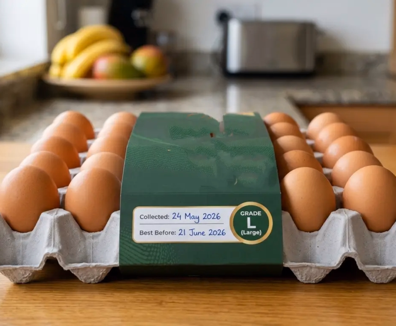
3. Freshness date on tray with internal quality guarantee: A tray label that states “Grade AA internal quality guaranteed within 7 days of packing date” — backed by the farm’s knowledge of its own HU tracking data — communicates the quality promise in terms the buyer can verify.
A farm that tracks Haugh units weekly, adjusts nutrition when quality declines, optimizes storage temperature to maintain the commercial window, and communicates that quality management system to buyers is not just selling eggs. It is selling assurance — the assurance that every tray will perform in the kitchen the way the last one did.
That assurance is the premium. The Haugh unit is the measurement that makes it credible.
Summary
Internal egg quality — Haugh units and yolk strength — is built during the 7–10 days of follicular development before ovulation, shaped by the nutritional status of the hen, and maintained or degraded by storage temperature after laying. It is the quality that consumers experience at the moment of use and that institutional buyers judge against for supply contract renewal.
The factors that depress internal quality below genetic potential are manageable: heat stress compensation through nutritional adjustment, antioxidant protection of the oocyte through vitamin E and selenium, blood spot reduction through vitamin K and vitamin C, oviduct secretory support through riboflavin and choline, and mycotoxin elimination through feed quality management.
The weekly break-out assessment protocol provides the data. The threshold triggers identify when intervention is needed. The supplementation protocol provides the intervention. The institutional quality communication framework closes the loop between what the farm produces and what the buyer verifiably receives.
An egg that breaks Grade AA tells the buyer to reorder. Make sure yours does.